
NCERT Class 9 Science Chapter The Fundamental Unit of Life is a gateway to the primary concept of biology—the Living Unit. Cells make up all living things, whether they are plants, animals, or microbes. Knowledge of the cell’s structure and functions is one of the ways to get a strong grip on biology. The chapter elaborates on the reasons why the cell is referred to as the basic structural and functional unit of life, thus making it a necessity for attaining academic success and gaining a good grasp of the concepts.
Through the Cell study, students are able to see the life processes happening at the microscopic level. Such topics as the interrelations of different basic Units of Life components that are responsible for the vital functions like respiration, nutrition, and reproduction are explained in the NCERT 9th Science chapter. Understanding the cell, one can see the clear distinction between unicellular and multicellular organisms as well as how complexity in living beings occurs.
In The Fundamental Unit of Life, Class 9 learners examine the historical aspect of the Cell, its categories, and its organizational structure. The chapter touches on significant concepts like the plasma membrane, nucleus, cytoplasm, and cell organelles, all presented in an uncomplicated and orderly fashion. A solid grasp of the basic Unit of Life becomes a factor for the success of the student in examinations and also a prerequisite for further studies in biology in the future.
The Microscopic Unit is not merely a concept of biology; it is a term that also refers to all the processes that living organisms carry on to grow, heal, and keep themselves alive. The subject is made more interesting and easier to comprehend through NCERT Grade 9 Science’s usage of drawing and real-world examples that provide students with a sound visualization of the cell’s architecture. Moreover, this chapter also explains the distinction between plant and animal cells, which is a major topic of assessment for students.
Table of Contents
Introduction to Cell – The Basic Unit of Life
A Cell is the tiniest living component that forms the entire structure of all life forms. One or more cells make up every plant, animal, and even microorganism. All life activities occur inside the cell; hence, it is referred to as the unit of life. In Class 9 Science NCERT, learning the Microscopic Unit is very much necessary, as it is the basis of biology. Without the Microscopic Unit study, it would be impossible to know how the living organisms grow, sustain, and reproduce.
The Cell carries out all other life activities, which are respiration, inhalation, elimination of waste, and procreation, amongst others. In the case of single-celled organisms such as Amoeba, respiration is done by one cell only, but in the case of multicelled organisms, the different cell types do their respective things together to keep the life going. The body is made more efficient by this division of work. By teaching about the Microscopic Unit, students can see how complex organisms are composed of small living units.
Cells are different from each other in all aspects, such as shape, size, and structure, according to the function performed. For instance, the structure of nerve cells is elongated to pass messages, whereas the structure of red blood cells is circular to enable oxygen transport to be more efficient. Despite these variations, each Microscopic Unit comprises the cell membrane, cytoplasm, and genetic material as the common constituents. The study of the cell gives students insights into the close link between structure and function, which is a key concept in biology.
The discovery of the Cell was a turning point in the study of life sciences. The advent of microscopes facilitated the observation of cells by scientists and the comprehension of their internal structure. The understanding of the cell is nowadays beneficial in medical, biotechnological, and genetic fields. For Class 9 students, the strong Microscopic Unit concept not only assists in getting good grades in the exams but also serves as a solid foundation for higher classes.
Discovery of Cell and Its Historical Background
The cell’s discovery was a milestone event, which changed the very basics of human perception of living forms thus the very concepts such as that of plant and animal anatomy through the microscope, etc., and so on. The English scientist Robert Hooke was the first person to discover the cell in 1665 when he looked at a very thin slice of cork under a microscope. He saw little box-like structures and called them “cells” because they reminded him of small monk’s rooms. This observation was the start of cell biology and the dominant view that living beings are made of tiny structural units.
It is true that Robert Hooke was the first person to discover cells, but the Microscopic Unit he saw were dead ones. In 1674 another scientist Anton van Leeuwenhoek took over with a more sophisticated microscope to view living cells. He explored the water in ponds and inferred etc. He called them “animalcules”. His finding provided again more convincing proof that cells are structures where the activities of life take place. This progress opened up further studies of the cells in detail.
Gradually, the Microscopic Units became less of a mystery thanks to the work of countless researchers. The year 1838 saw the birth of the plant cell theory with the disclosure that all plants are made up of cells by Matthias Schleiden; the next year brought the revelation that animals are also cell-made from Theodor Schwann’s research. They were the trio who paved the way for the development of cell theory through their research and proclamations that cells are the tiny things that make up all living things. Later on, Rudolf Virchow went a step further to proclaim that the cycle of life starts with division of cells, thereby reinforcing the whole cell concept once again.

The Cell’s discovery, with its historical background, provided strong evidence for the understanding of the life cycle of living organisms in terms of growing, developing, and reproducing. This knowledge is fundamental not only for biology but also for medicine and biotechnology. For ninth graders, the cell’s discovery recount makes it easier to pinpoint the roots of biological concepts, thus facilitating a more comprehensive grasp of life sciences.
What is a Cell? Definition and Characteristics
A cell is the basic unit of life that has both structure and function. All living organisms, be it plants, animals, or microorganisms, are made of cells, either one or many. Inside the cell, all the life activities such as growth, respiration, nutrition, and reproduction occur. This is the reason why the Microscopic Unit is regarded as the basic building block of life and is an essential topic in Class 9 Biology.
The cell possesses the features of a living unit. It can grow, divide, and respond to the environment. Through the process of metabolism, the Microscopic Unit performs all the chemical reactions that are necessary for life. Some organisms are unicellular, like Amoeba, while others are made up of millions of cells that work together. All these characteristics point out that the cell is not just a structure but a living and active unit.
Depending on the body part where they are located, cells are different in size, shape, and function. For instance, the muscle cells are long and elastic, so they can easily stretch and help in movement, while the nerve cells are built for sending signals. Nevertheless, all Microscopic Unit have some common features such as a cell membrane, cytoplasm, and DNA. By studying the cell, students are able to see the relationship between the structure and the function of living organisms.
The knowledge of the cell and its properties is always a necessity in the study of biology. Such knowledge is the basis for a student’s future learning in higher classes of tissues, organs, and organ systems. For students in Grade 9, a thorough understanding of a Microscopic Unit, its structure, and functions makes the subject of biology easier and thus helps in securing a good mark in the exams. Besides, the concept of the cell also has practical applications in fields like medicine, genetics, and biotechnology.
Types of Cell: Unicellular and Multicellular Organisms
Organisms are grouped into various categories according to the cell count in their bodies. The basic unit is the cell, and thus, organisms are split into two classes: unicellular and multicellular organisms. Unicellular constitute a single cell, whereas multicellular organisms consist of numerous cells working in unison. Learning these classifications of cells helps students comprehend the architecture of living beings and the operation of life processes.
In the case of unicellular organisms, the lone Microscopic Unit carries out every single life function, including respiration, nutrition, movement, and reproduction. Some of the unicellular creatures are Amoeba, Paramecium, and bacteria. In most cases, these organisms are very tiny and are able to live on their own. They are single-cell organisms, yet they are full-fledged living beings that can perform all the vital functions of life.
On the other hand, multicellular organisms are the result of a mix of cells with different specializations. Each Cell in the organism performs a different function – for example, muscle cells are responsible for movement, nerve cells are responsible for transmitting signals, and blood cells are responsible for transportation. Examples of multicellular organisms are: plants, animals, and humans. The division of labour among the Microscopic Unit makes the organism more efficient and complex.
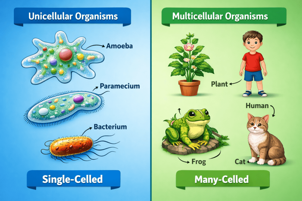
The study of unicellular and multicellular organisms aids the student’s knowledge about the Cell’s role in supporting the complex life forms. It also illustrates the tissue, organ, and organ system development in multicellular organisms through cell cooperation. For Class 9 students, this subject is a necessity for grasping advanced biological concepts and for being successful in tests. The primary understanding of cell classification is the groundwork for further education in biology.
Cell Shape, Size, and Number
A cell’s shape, size, and number can change according to its function and the organism’s type. While all living organisms consist of cells, their structures vary to some extent. A cell’s shape is determined by the nature of its work. For instance, nerve cells are elongated and branched to conduct signals rapidly, whereas red blood cells are circular in shape to transport oxygen easily through blood vessels.
The size of a cell varies greatly among different organisms. Some cells are so small that they can only be seen with the help of a microscope, such as bacterial cells, while others are large enough to be seen without any aid, like the egg of an ostrich. Generally, cell size in plants and animals is kept very small, which allows oxygen and nutrients to be quickly exchanged with the surrounding medium. A smaller cell size advantageously makes for better substance exchange.
The number of cells in an organism depends on whether it is of the unicellular or multicellular type. In the case of unicellular organisms, one cell takes care of all the functions of life, while in multicellular organisms, there are millions of cells that cooperate to produce tissues, organs, and systems. Each cell in a multicellular organism is designated to perform one special function, which in turn makes the organism more effective.
The study of the shape, size, and number of cells enables the students to understand how the structure relates to the function in living organisms. This unit shows the different appearances of cells and the way their design assists them in performing their part in the body. For students of the ninth grade, this understanding is essential in the formation of solid biological concepts and the accurate answering of examination questions. The students are also ready for the higher topics in biology through the clear knowledge of cell variations.
Cell Structure Explained with Diagram
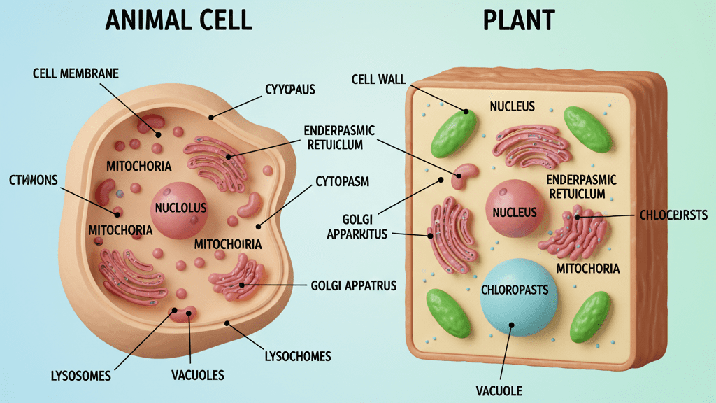
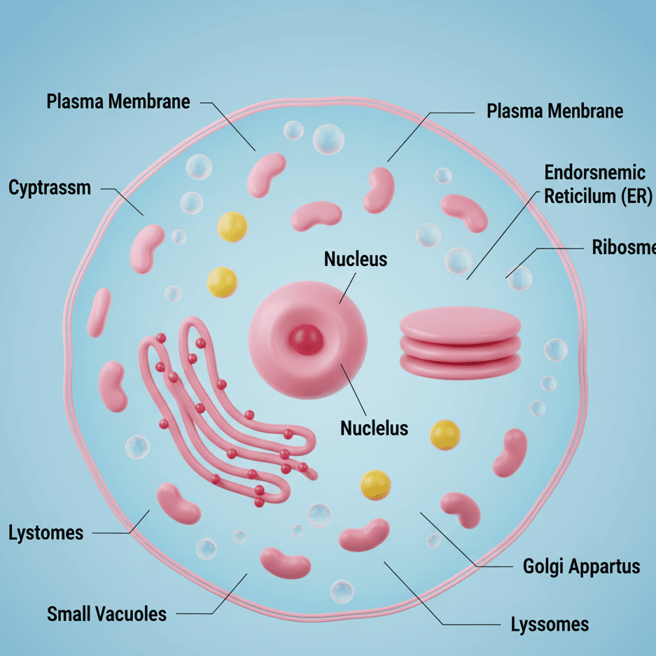
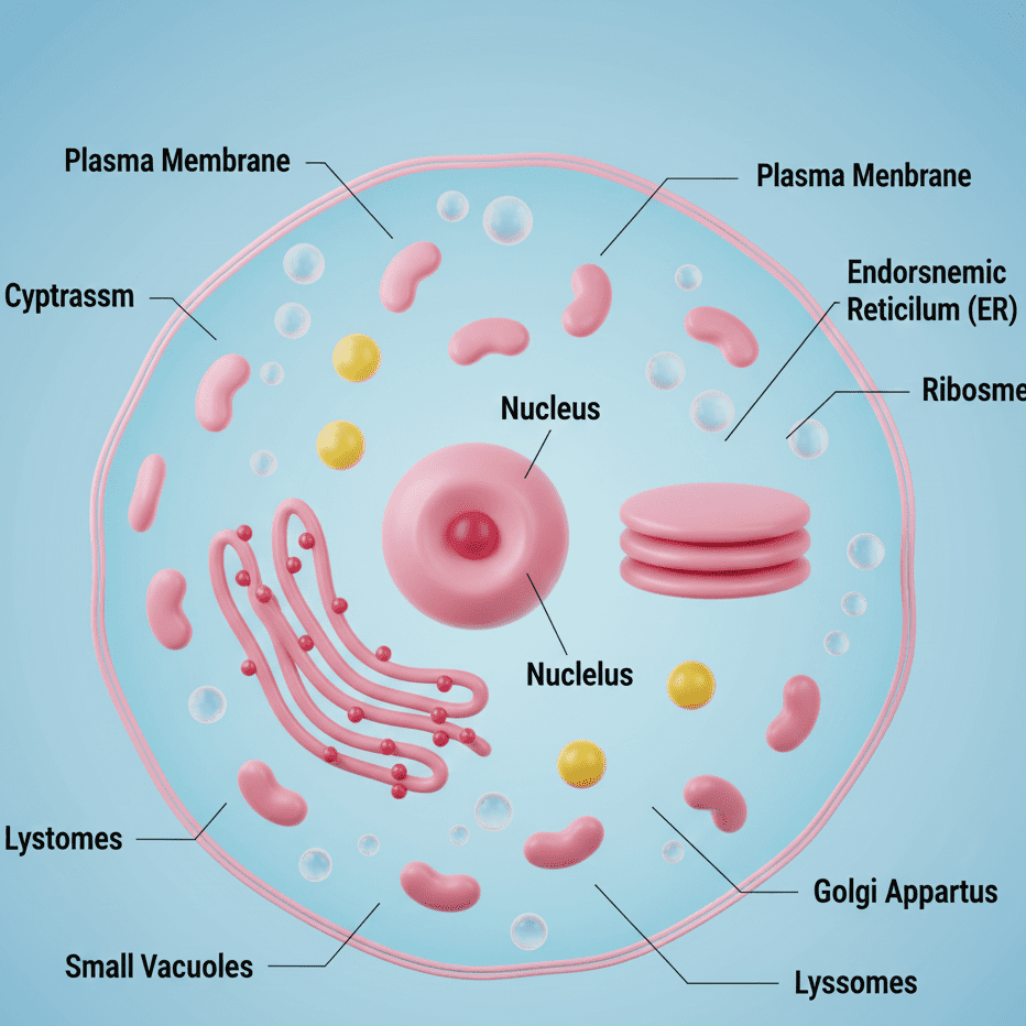
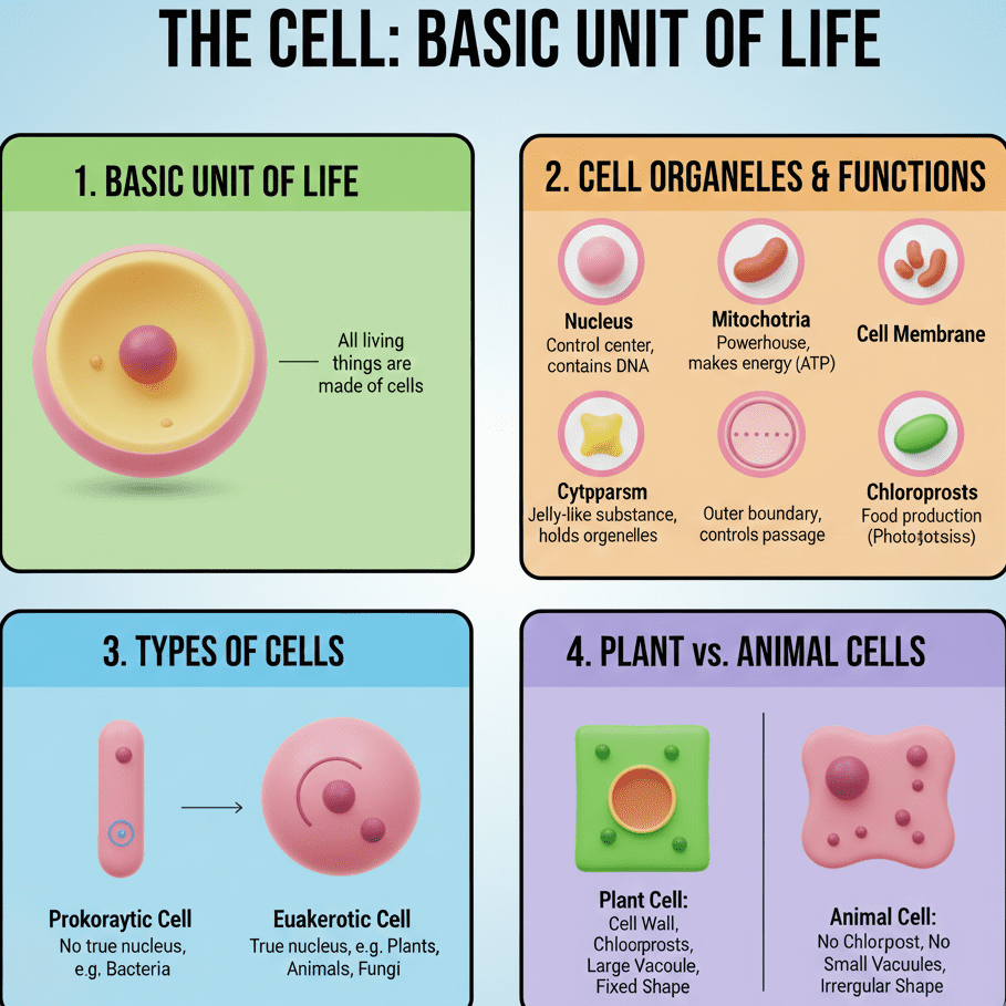
The body of a Cell reveals to us the secrets of the living world on a microscopic level. All living things have their own cells, and they all share the same prevailing features; the cell’s anatomy is the basis of the cell’s activities, which are essential for life. Throughout the 9th-grade class, especially in the NCERT science syllabus, one can learn the anatomy of the cell well through the use of a diagram, and this will not only be a thing of visualizing but also remembering the cell’s parts and their functions. A cell diagram with its parts marked out is a very significant part of the exam preparation.
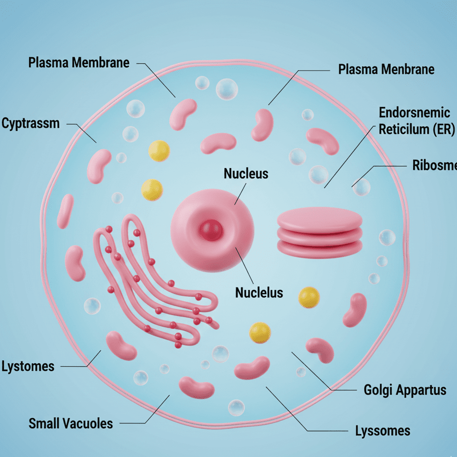
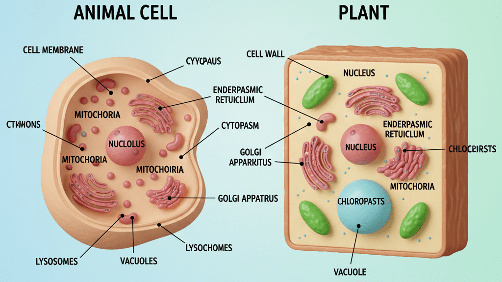
A cell that is conventional in nature comprises three primary structures: the cell membrane, cytoplasm, and nucleus. The outermost layer of the cell is the cell membrane; it carries out the function of being the controlling factor of substances entering and leaving the cell. The cytoplasm is the watery and thick substance of the cell where most of the chemical reactions occur. The nucleus is the part that contains the genetic material, and it directs all the cell’s activities. The three parts are interdependent, and they are all essential for the cell’s life.
The cell is not only composed of the fundamental parts but also includes a variety of cell organelles, like mitochondria, endoplasmic reticulum, Golgi apparatus, ribosomes, and lysosomes. Succeeding the life of the cell is the specific function of each organelle; for instance, mitochondria generate energy while lysosomes eliminate waste materials. To illustrate, diagrams facilitate students’ comprehension of cell organelles’ location and function.
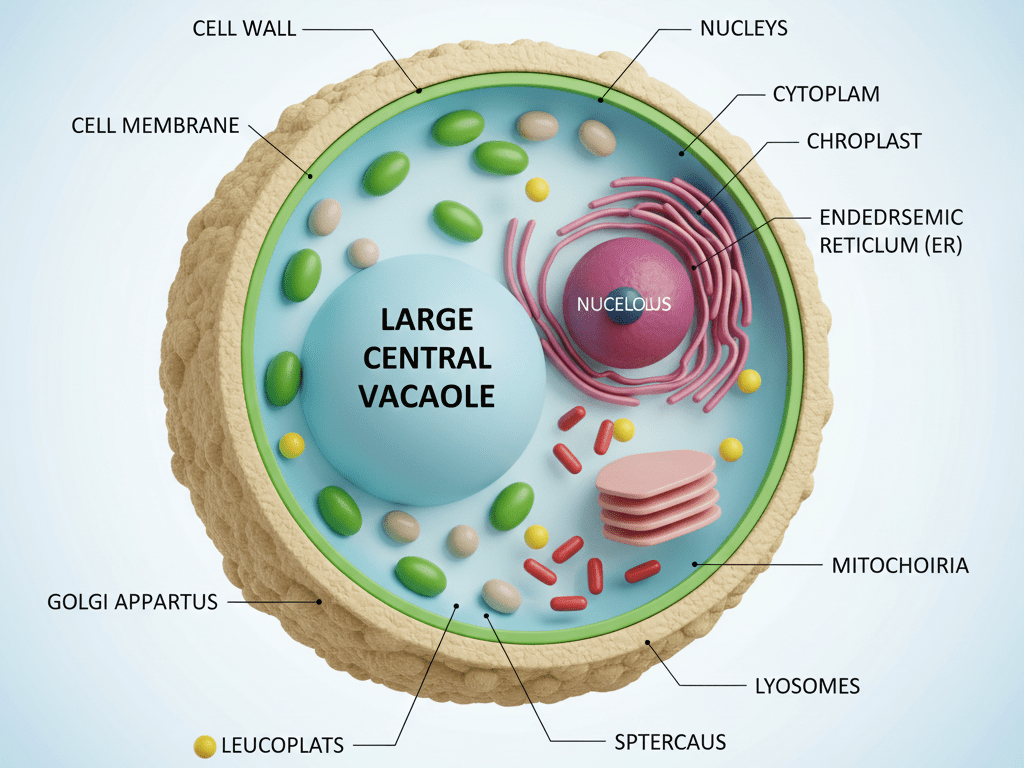
It is through diagrams that students learn and retain better the cell’s structure. Quite literally, the learning in this case is visual; the student sees how parts of a cell are organized and how they interact. For the class 9th students, this topic is of great importance in terms of answering diagram-based questions and giving long answers in exams. A profound knowledge of the cell’s structure indeed is the first step in facilitating one’s understanding of higher biology study areas.
Plasma Membrane: Structure and Functions of Cell Membrane
The plasma membrane, or cell membrane, is the outermost layer of the cell. It distinguishes the cell from its environment and safeguards the cell’s internal parts. The plasma membrane is thin, flexible, and living in nature, which enables it to perform important functions. Comprehending the structure of the plasma membrane is a necessity for Class 9 pupils because it is central to cell life.
The plasma membrane structure of a cell consists of specifically arranged lipids and proteins. This arrangement is commonly described by the fluid-mosaic model, wherein the proteins are embedded in a layer of lipids. The unique structure gives rise to the selectivity of the plasma membrane to permeability, meaning it allows the passage of some substances and at the same time restricts others. This property is essential for the cell’s internal environment to be constant.
Controlling the movement of materials in and out of the cell is one of the foremost functions of the plasma membrane in a cell. Processes like diffusion, osmosis, and active transport take place across the plasma membrane. These processes enable the cell to absorb nutrients, excrete waste, and keep substances at the right concentration. Absence of a functional plasma membrane would lead to the cell’s death.
In addition, the plasma membrane helps the Cell to shape up and provides a barrier against toxic materials. In multicellular organisms, the cell membrane provides a medium through which cells can send messages to and receive signals from one another; thus, they can function in an organized manner. Having a solid understanding of the plasma membrane and its functions is a major help for Class 9 students in answering conceptual and application-based questions in exams. Moreover, this topic serves as a stepping stone for understanding intricate biological processes in higher classes.
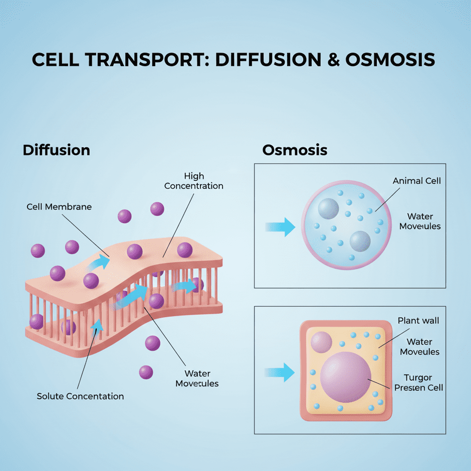
Diffusion and Osmosis in Cell
Diffusion and osmosis are two major processes that underlie the existence of any living cell. The processes in turn are a medium through which the cell exchanges substances with its environment. The process of diffusion refers to the transfer of particles from a place where they are more concentrated to a place where they exist in smaller quantities, and osmosis is the water movement across a selectively permeable membrane. The comprehension of both diffusion and osmosis is a prerequisite for understanding the cell’s material entering and exiting.
Diffusion is a process that facilitates the movement of gases such as oxygen and carbon dioxide across the cell membrane in a Cell. No energy is used in the process and it happens naturally due to concentration gradients. For instance, oxygen is absorbed by the cell for respiration while carbon dioxide is expelled as a waste product. Diffusion is a means of enabling the cell to remain in balance with its environment and at the same time, it is a part of the necessary life activities of the cell.
Osmosis is a unique diffusion case where only water molecules go in or out of a Cell through the plasma membrane. If a cell is put in hypotonic solution, the water comes into the cell which causes the cell to swell. On the contrary, in hypertonic solution, the water inside the cell is drawn out which results in cell shrinkage. Such changes provide evidence that osmosis is one of the main factors that regulate the water content of the cell and its shape.
Diffusion and osmosis are among the main factors that contribute to the survival of the internal environment of a cell. These processes are the ones that facilitate the inward transfer of nutrients, excretion of waste, and doing so in a way that the concentration of substances inside the cell remains proper. For Class 9 students, taking a step back and looking at diffusion and osmosis, the processes must be seen as important background for dealing with concepts and practical questions in exams. A good understanding of these topics is also a door opened for a good understanding of biological processes in higher classes.
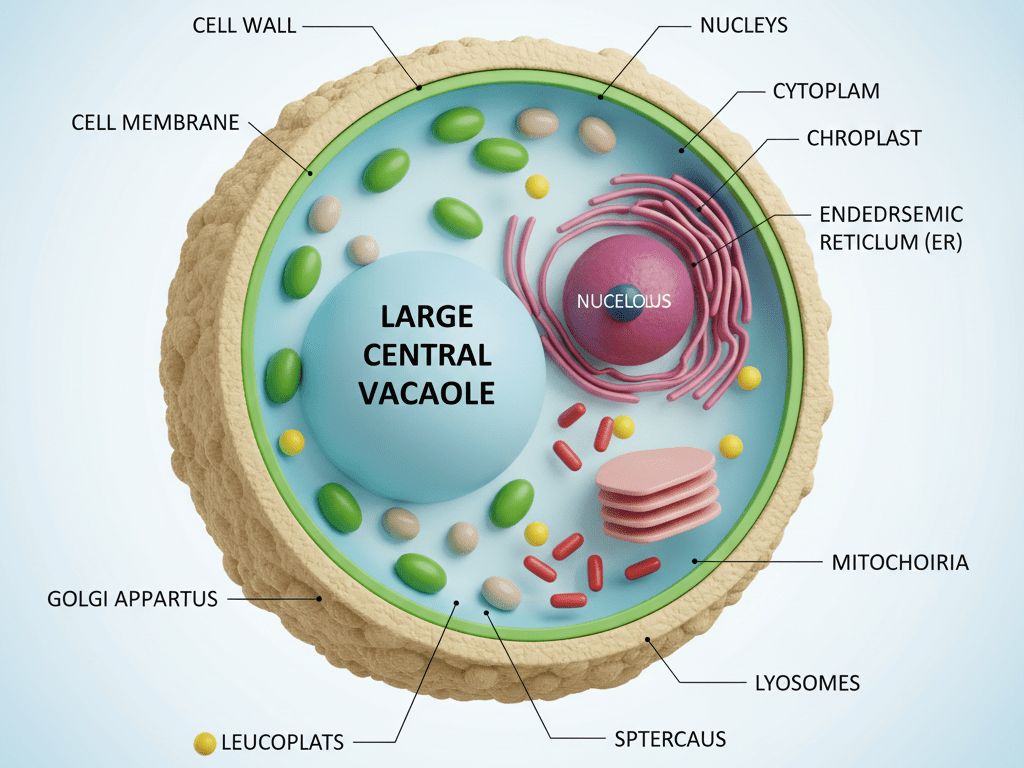
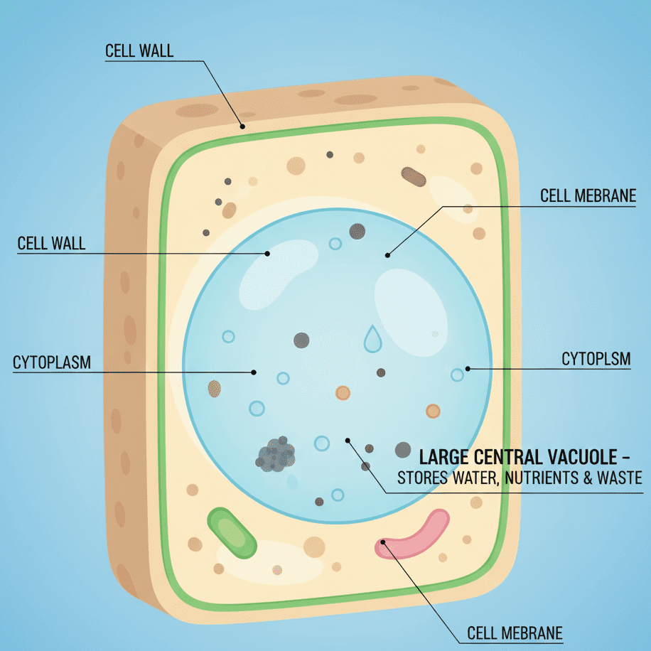
Cell Wall: Structure and Role in Plant Cell
The cell wall is a remarkable boundary layer that is present in every plant cell. It is a tough layer that lies outside the plasma membrane and serves to safeguard and bolster the cell. The cell wall is, unlike the membrane, dead and non-flexible. The study of the structure of the cell wall reveals to the students that the plant cell has a fixed shape and is, therefore, stronger than the animal cell.
The makeup of the cell wall or plant cell is primarily cellulose, along with hemi-cellulose and pectin, which are the minor components. This powerful build-up is responsible for the cell wall’s rigidity and toughness. In fact, the cell wall is fully permeable, that is to say, it allows substances to pass through easily. It is this very property that facilitates the movement of water, gases, and nutrients into and out of the cell.
The cell wall is the first line of defence in a plant cell against mechanical damage, and its main function is to keep the cell alive and its character. It also acts like a sponge in that it prevents the cell from getting too big and eventually bursting when pulling in excess water, a process called osmosis. The plant, with its wall, becomes a tall, sturdy statue. Plant cells would not be able to maintain their structure and stability without a strong cell wall.
Grasping the function of the cell wall is a must in order to recognize the differences between plant and animal cells. This topic is very helpful for class nine students in their preparation for examination, as they will be able to answer both conceptual and comparison-type questions. The cell wall also reveals many processes related to plants, such as their growth and absorption of water, to be understood. In this way, a clear understanding of the subject matter is going to be a strong step towards advanced studies in plant biology.: Structure and Role in Plant Cell
Cytoplasm: Internal Environment of the Cell
Cytoplasm is a thick and viscous liquid that is found in all cells; it is located inside the cell membrane and outside the nucleus. It is the part of the cell that can be considered “alive,” since a great deal of the life processes take place in the cytoplasm. The cytoplasm is made up of water, salts, proteins, as well as a variety of cell organelles. Learning about the cytoplasm gives students an idea of how the different parts of the cell work together to support life.
Cytoplasm in a cell is the means in which all the cell’s organelles are held such as the mitochondria, ribosomes, the endoplasmic reticulum, and the Golgi apparatus. The organelles are a part of the cytoplasm and are engaged in their specific functions. Moreover, the cytoplasm is the facilitator in the movement of materials within the cell, thus contributing to the proper functioning of the cell.
The cytoplasm of a Cell is the site of many important chemical reactions. The area of protein synthesis, metabolism, and enzyme activity is the cytoplasm. The cytoplasm is the place where these reactions get the necessary environment for their happening in an efficient manner. Without the cytoplasm, the cell would be dead, as it would not be able to perform its vital life activities.
The cytoplasm is a vital player in the Cell’s maintenance of the shape and the cellular organization on the inside. It is the cytoplasm that spreads and supplies the different cell organelles with nutrients, oxygen, and energy. For Class 9 students, having knowledge of the cytoplasm is a must for both short-answer and long-answer questions during examinations. Not only is this topic clearing up a strong base for learning advanced cellular processes in higher grades when it is understood well.
Nucleus: Control Center of the Cell
The nucleus is a key component of the Cell, and it is often referred to as the control center of the cell. Usually, it is found in the middle and is covered by a nuclear membrane. The nucleus has the genetic material that regulates the cell’s growth, development, and metabolism. Learning about the nucleus’ functions allow students to comprehend how cells are regulated and organized.
Chromosomes and the nucleolus are some of the structures within the nucleus of a Cell. Chromosomes bear the genes, which are the traits inherited from the ancestors. The nucleolus is active in ribosome formation. The various functions of the nucleus make it indispensable in controlling cell actions and in the production of proteins.
The nucleus is a decisive factor in cell division and reproduction in a Cell. In the process of cell division, the nucleus makes sure that the genetic material is equally divided among the new cells. Additionally, it regulates gene expression and so controls a variety of metabolic activities. The cell would not be able to carry out its normal functions and would not be able to divide without a nucleus.
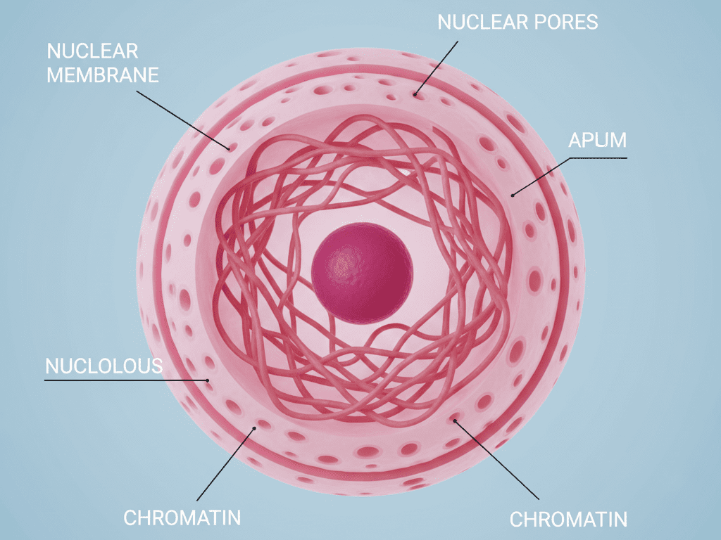
The nucleus is the control center of the Cell, and this is why Class 9 students need to learn it well. This area of study bridges the gaps in heredity, growth, and development in the organism. Besides, since a clear understanding of the nucleus is a prerequisite for accurate answers in exams, it thus prepares the students for the subsequent topics of genetics and cell division in higher classes.
Mitochondria: Powerhouse of the Cell
Mitochondria are the cells’ powerhouses, which means they are vital organelles that practically every cell has. They produce the energy needed for all the life processes going on in the cell. The energy that mitochondria generate is kept in the form of ATP, which is commonly referred to as the cell’s energy currency. The comprehension of mitochondrial function can lead the students to the conclusion that cells are capable of getting a boost in energy, which is necessary for the performance of their vital functions.
The analysis of the cell mitochondria structure reveals a peculiarity and an architectonic order. The structure is characterised by a double membrane, outer and inner membranes. The inner membrane has folds called cristae, which greatly enhance the area for the energy production. A fluid-filled space called the matrix, where significant chemical changes happen, is found inside the mitochondria.
In a cell, mitochondria are involved in and are the regulators of the cellular respiration process. The breakdown of food substances, for instance, glucose, takes place with the intake of oxygen to the point where energy is released. This energy is then converted to ATP, which is the powerhouse for the cell’s activities, such as growth, movement, repair, and many others. Muscle cells that need more power, for instance, house a greater number of mitochondria.
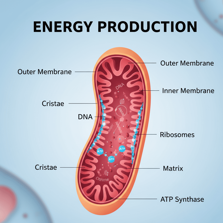
The inquiry into why mitochondria are termed the powerhouse of the cell is very significant for the understanding of class 9 students. This issue is of great help in the elucidation of both conceptual and application-based questions that are usually asked in exams. The comprehension of mitochondria also serves as a solid basis for the more advanced topics like respiration and metabolism in the later classes. A sound knowledge of this topic makes the study of biology less difficult and, at the same time, more enjoyable.
Endoplasmic Reticulum (ER): Transport System of the Cell
The endoplasmic reticulum (ER) is one of the major organelles and is present in every eukaryotic cell. It serves as a transport system that facilitates the distribution of substances between cell parts. The ER is a network of membrane-enclosed tubes and sacs that are in close proximity to the nucleus. By knowing the ER’s function, students can recognize how different compounds are very efficiently moved inside the cell.
In a cell, the endoplasmic reticulum is divided into two principal categories: the rough endoplasmic reticulum (RER) and the smooth endoplasmic reticulum (SER). The rough ER is equipped with ribosomes on its surface and is predominantly engaged in the production of proteins. The smooth ER, on the other hand, lacks ribosomes and participates in the manufacturing of lipids and fats. These two types of ER are not only different but also equally significant in their respective functions for the cell.
The endoplasmic reticulum is one of the major components of the cell that determines its internal structure. To a great extent, it is responsible for the cell’s surface area where the chemical reactions occur, besides transporting the proteins and lipids to the specific sites in the cell. Also, in some cells, the smooth ER is involved in the detoxification process of the harmful substances. Without the ER, the cell would be in a tough position managing the production and circulation of the vital materials.
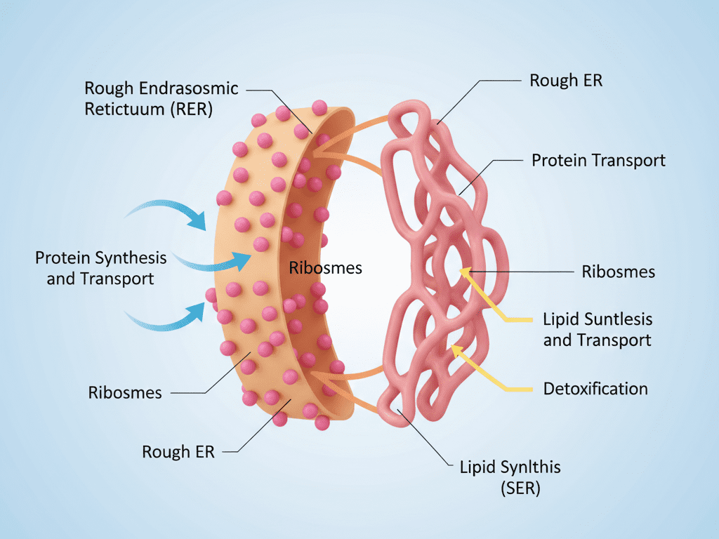
Cells have to rely on the endoplasmic reticulum as the transport system of the cell. This concept is very important for Grade 9 learners. It aids in unfolding the story of cell organization and cell functioning. Besides being a fundamental concept for students, a clear understanding of the ER will also help them answer questions coming from this topic during the exams and keep them ready to encounter more challenging biological knowledge later on in higher classes.
Ribosomes: Protein Factory of the Cell
Ribosomes, although small, are crucial structures found in all living cells. They are commonly referred to as the cell’s protein factory because they perform the primary function of producing proteins. The cell’s proteins are necessary for its metabolism, growth, and repair. If students comprehend the function of ribosomes, then they can easily learn the process through which cells create their own materials for survival.
Ribosomes may either be found in the cytoplasm or be attached to the rough endoplasmic reticulum (ER) in a cell. The rough ER-bound ribosomes participate in the production of proteins that are either sent to other regions of the cell or outside the cell. In contrast, free ribosomes generate proteins for use within the cell itself. This indicates that ribosomes operate very efficiently according to the cell’s demands.
In a cell, ribosomes use the genetic code obtained from the nucleus to carry out protein synthesis. Protein synthesis is a vital process of all living organisms and is unavoidable for life activities. The newly formed proteins have various functions as they become enzymes, hormones, and structural elements. If there were no ribosomes, the cell would be unable to perform functions such as growth, repair, or other critical metabolic functions.
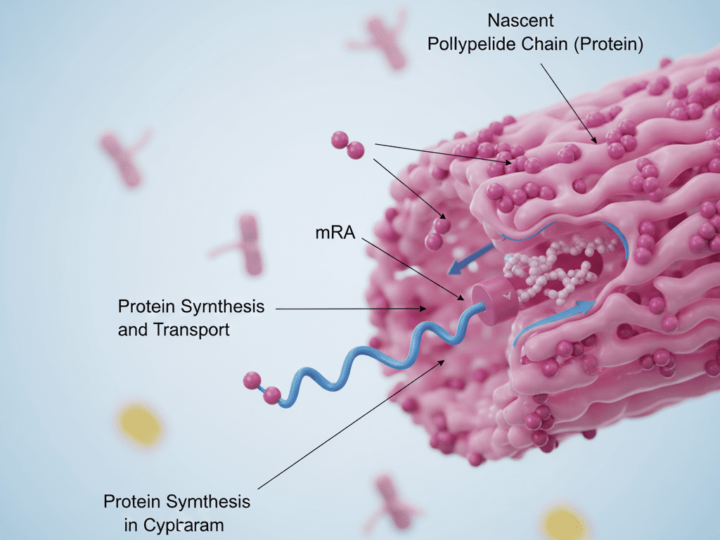
Understanding the reason behind ribosomes being termed the protein factory of the cell is very fruitful for the 9th-grade students. The topic not only helps students to tackle conceptual questions in examinations but also lays a strong groundwork for advanced biology topics such as gene expression and metabolism. A clear understanding of ribosomes makes the study of cell biology easier and more interesting.
Golgi Apparatus: Packaging and Secretion in Cell
The Golgi apparatus is a cell organelle that is vital, and its main role is to provide a packaging and secretion process for cells. It performs this function like a cell’s post office, by receiving proteins and lipids from the endoplasmic reticulum. After receiving these materials, the Golgi apparatus alters, categorizes, and prepares them for delivery to different places within the cell or outside it. The importance of the Golgi apparatus in cell functioning helps students in recognizing the efficiency of cells in the management and transportation of substances.
The Golgi apparatus’ structure in a cell is made up of a set of flattened and membrane-bound sacs known as cisternae. These sacs are piled up one over the other and linked with tiny vesicles. The Golgi apparatus encapsulates the materials generated in the cell into vesicles. This well-organized arrangement allows the cell to rightly process and transport both the proteins and the lipids.
A cell’s Golgi apparatus is mainly engaged in the trio of processes that include packing, storing, and discharging of substances. The proteins and lipids synthesized in the endoplasmic reticulum are dispatched to the Golgi apparatus, where they undergo modification and are eventually packed into vesicles. The vesicles then transport the substances to their respective locations. This whole procedure is what keeps the cell running in a smooth and efficient manner.
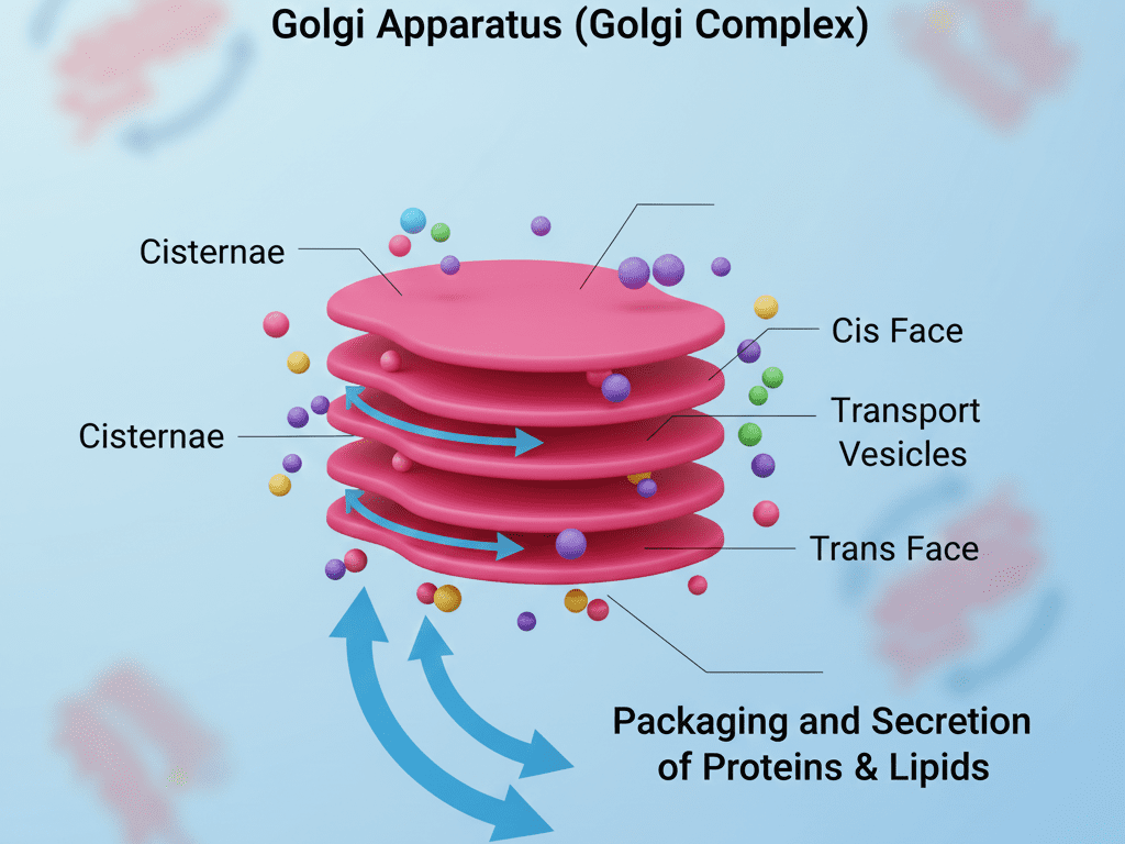
Class 9 students must view the Golgi apparatus as the Cell’s orchestrated and expelled system. This notion is one of the key areas in the study of cell organelles and their functions to satisfy exam questions. The students’ grasp of the Golgi apparatus will eventually guide them through the more challenging biological concepts in later classes such as secretion, transport, and cellular organization.
Lysosomes: Suicide Bags of the Cell
Lysosomes, which are organelles with membranes, are located in the cell and are referred to as the “suicide bags of the cell.” They are filled with very strong enzymes, called digestive enzymes, that are necessary for breaking down waste materials, damaged organelles, and foreign particles. The students’ comprehension of lysosomes will help them to get the idea of how cells maintain their cleanliness and health by purging the noxious materials.
The construction of lysosomes in a Cell is plain but very efficient. They are tiny, round, and full of sloppy enzymes that are very active in the acidic atmosphere. These enzymes can digest proteins, fats, and carbohydrates. When a cell’s life cycle is over, or it gets impaired, lysosomes set free their enzymes and digest the whole cell, hence they are also called suicide bags of the cell.
Lysosomes are vital in the process of balancing the cell’s internal environment. They are part of intracellular digestion, defense against harmful microorganisms, and recycling of cell parts. This recycling process consumes raw materials which the cell can reuse. If lysosomes were not there, the cell would be in trouble because the waste would accumulate and harm the cell eventually.
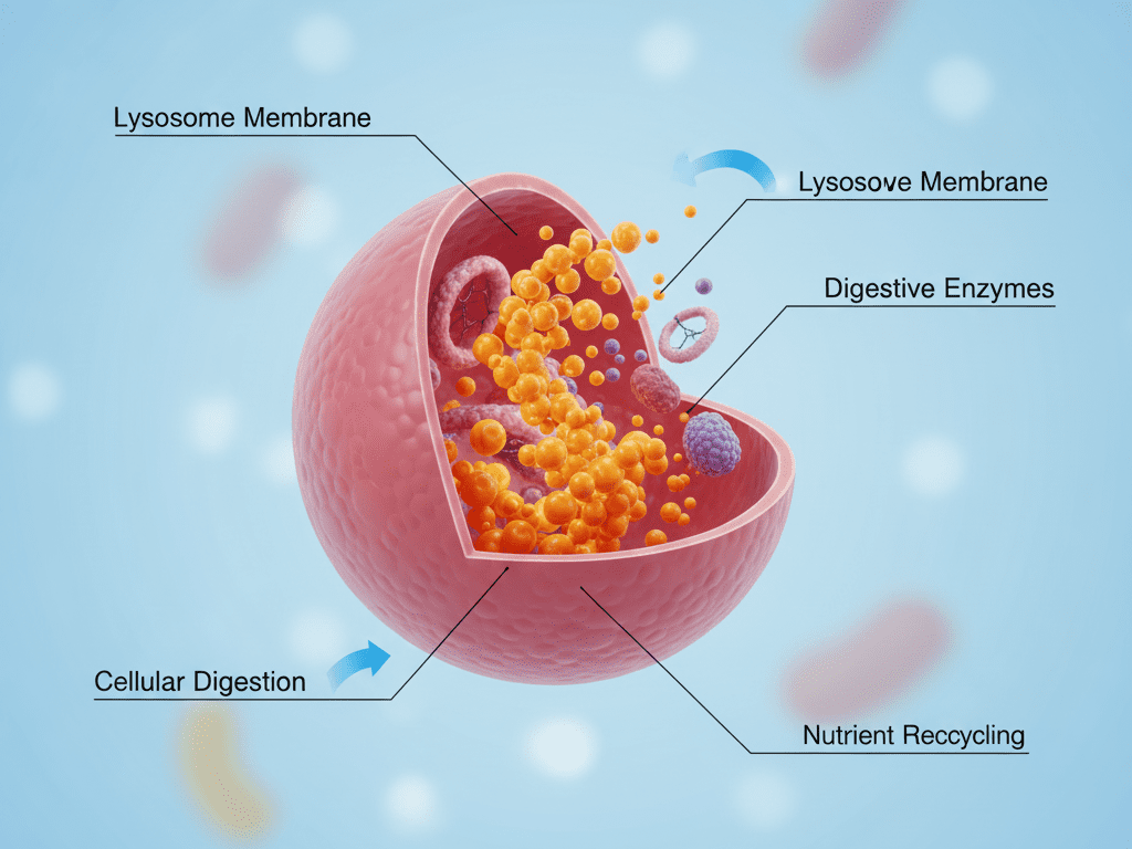
The argument of why lysosomes are called the suicide bags of the Cell is quite crucial for the students of Class 9. This topic is helpful in promoting the students’ comprehension of conceptual and application-based questions in examinations. Knowledge about lysosomes also provides a solid basis for advanced topics like cell maintenance and immunity in higher grades. A clear comprehension of this organelle not only makes cell biology easy but also exciting.
Vacuoles: Storage and Regulation in the Cell
Vacuoles, the cellular structure found in the cell, are the main areas that are responsible for storage and are therefore known as the storage sites. They can be filled with water, food, salts, and waste. In the case of plant cells, vacuoles are generally large and thus cover a large part of the cell, while in animal cells, vacuoles are smaller or sometimes not even present. Knowing about vacuoles is an easy way for students to learn how cells handle the important stuff.
The vacuole structure of a cell is not complicated; rather, it consists of a wall called the tonoplast, which encloses the material to be stored. The liquid located inside the vacuole is referred to as cell sap. The construction of the vacuole enables it to hold considerable amounts of things without putting other cell processes on hold. The tonoplast also plays a role in controlling the entrance and exit of substances into and out of the vacuole.
Vacuoles are organic components that play a crucial part in the cell’s internal balance maintenance. They assist in plant cell turgidity and thus firmness owing to the pressure developed by the stored water inside the vacuole, supporting the plant. In addition, vacuoles facilitate waste disposal by storing undesirable substances and keeping them away from the cell’s metabolic activities. This implies that vacuoles are key players for cells to perform their functions properly.
For class nine pupils, it is very important to acknowledge the role of vacuoles in the storage and regulation of the cell. This subject matter is very much interlinked with the question “to explain plant cell structure and functions” in students’ examination scripts. Comprehension of vacuoles will also be a good preparatory ground for learning plant physiology and cell biology in high school students’ future courses.
Plastids: Chloroplast, Chromoplast, and Leucoplast
Plastids are one of the main organelles present in the plant cell. They are responsible for green color, which sometimes indicates the storage of the food, also known as photosynthesis. The plastids are classified into three major types: chloroplasts, chromoplasts, and leucoplasts. To have a basic knowledge of plastids is necessary for students, as those organelles are the central players in determining the structure and function of plant cells.
The green plastid, the chloroplast, which contains chlorophyll. chlorophyll is the green pigment that absorbs light in all wavelengths except green. Chlorophyll is found in chloroplasts, which gives plant green color. Besides saying ‘the production of food by the cell will not take place without the chloroplasts, we can also say that the energy produced by the cell will be diverted to the plant, and so positive growth and development will occur.
Like photosynthesis, the role of chromoplasts is opposite to that of chloroplasts. They are the colored plastids in some plant tissues. They color the fruits, flowers, and some leaves in red, orange, and yellow. In a plant Cell, the colored plastids help attract insects for pollination and animals for seed dispersal. Chromoplasts do not directly help the photosynthesis process but contribute indirectly to the plant’s survival and reproduction.
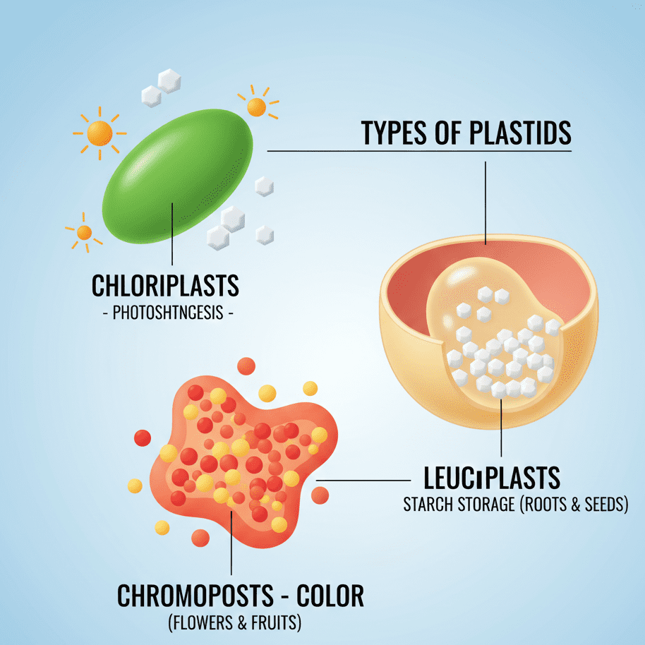
Leucoplasts are the plastids that are devoid of color and are found in those parts of the plant that do not carry out photosynthesis, like roots, seeds, and storage organs. They are the main ones to store starch, proteins, and oils. Leucoplasts help the plant store essential nutrients for later use, which is crucial for the growth and development of the plant. A clear understanding of plastids helps Class 9 students answer questions related to the structure and functions of plant cells and forms a strong foundation for higher-level biology.
Difference Between Plant Cell and Animal Cell
Plant cells and animal cells are living’s basic units in both structural and functional aspects, but also have some important distinctions. A Cell is the smallest living unit of the Plantae and Animalia kingdoms, but plant cells possess additional structures like the cell wall and chloroplasts, which are not found in animals. Learning these dissimilarities assists the students in knowing how, by means of specific functions, plant and animal cells work efficiently.
One of the most pronounced differences between a cell was the presence of a rigid cell wall in plant cells, which gives the cells shape and support, while the only feature of an animal cell was a flexible plasma membrane. Plant cells also have large central vacuoles for the storage of water and nutrients, whereas vacuoles in animal cells are small or temporary. The differences in structure have an impact on the functions of cells in plants and animals.
Yet another important difference in a Cell is the presence of plastids in plant cells. The presence of chloroplasts in plant cells allows the cells to gain energy directly from the sun through photosynthesis, while animal cells that lack chloroplasts must consume food for energy. In addition, plant cells are generally rectangular or polygonal in shape, whereas animal cells have a more round or irregular shape. These differences in morphology help the individual cell types to execute their respective roles efficiently.
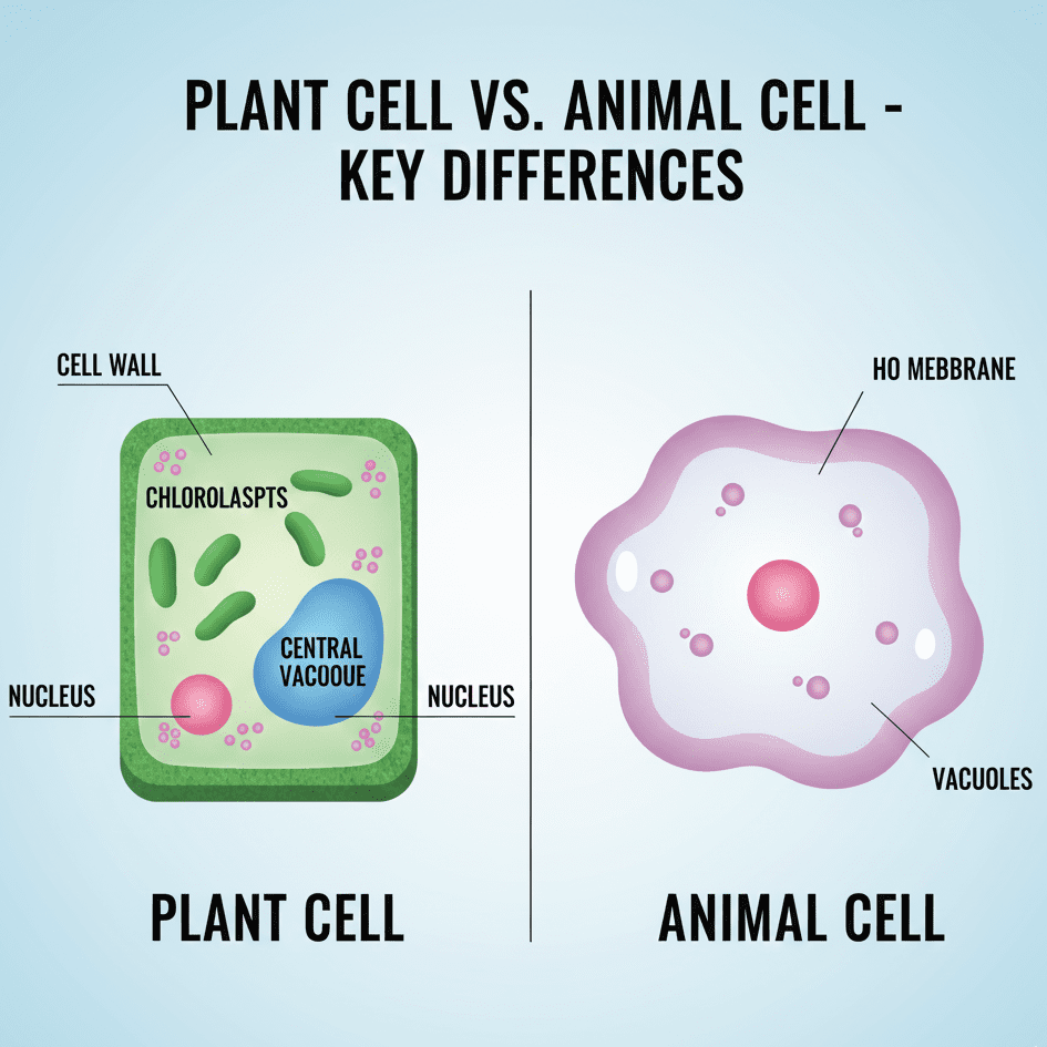
It is paramount for Class 9 students to learn the differences between plant and animal cells. The information acquired is useful in answering exam questions, making labeled diagrams, and fortifying biology at higher levels. Further, through these differences, students also realize that the structure of a Cell is intimately related to its function in living organisms, which makes biology easier to comprehend and more interesting.
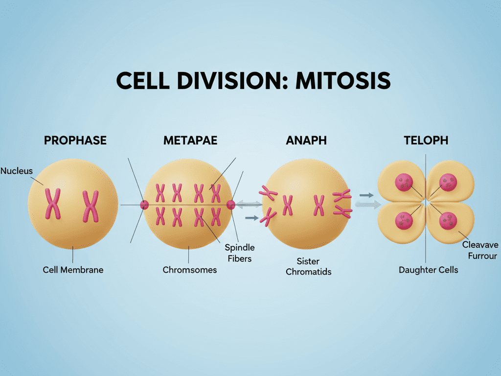
Cell Division – Introduction and Importance
Cell division is a term used to describe the cell’s own reproduction process, which results in two or more new cells. The process is crucial for the life cycle of organisms since it enables growth, repair, and maintenance. For that reason, cell division is at the heart of any living thing’s life cycle. The subject of cell division makes it easier for 9th-grade students to comprehend life’s continuity at the microscopic level.
There are generally two types of cell division in the life of organisms: mitosis and meiosis. In the case of mitosis, only one individual cell contributes its part and is eventually replicated into two identical daughter cells, thereby assisting the organism in its growth and repair activities. Whereas in meiosis, a single progenitor cell goes through a series of divisions and ultimately ends up producing four gametes, for instance, sperm and eggs, each of which carries half of the original cell’s chromosomes. It is through the learning of these different divisions that the students can realize how much cell division is involved in reproduction and survival.
Cell division plays an important role in keeping the chromosome number of a cell stable. When mitosis occurs, the genetic material gets an equal distribution and therefore arrives at the daughter cells unaltered. This is very important for ensuring that tissues and organs perform their respective functions without any interruption. In the case of improper cell division, cells may be harmed, and that, in turn, can lead to the organism’s growth, development, and health issues.
Being aware of the process of cell division and its necessity is a must for pupils in grade nine. This particular theme opens up the discussion of how organisms develop, replace tissues, and eventually. An unclouded understanding of cell division also enables students to progress through the genetics, heredity, and molecular biology sub-disciplines of biology. A Cell’s division and its necessity are thus the key to unlocking the secrets of biology and preparing students for more advanced studies.
Importance of Cells in Living Organisms
Cellular organisms are composed of cells, which are the actual microscopic living units. One could say that life is the living unit of cells. As such, it is the cell that is the building block of organisms. Therefore, the students should comprehend the importance of the cell to understand why it is the building block of life.
In organisms, cells assist in performing necessary life processes in an efficient manner. In the case of unicellular organisms, the single cell is responsible for all the functions vital for survival. However, in multicellular organisms, specialized cells cooperate to develop tissues, organs, and organ systems. This cell cooperation ensures that every single activity, whether it be blood flow, food processing, or breathing, is conducted most effectively, resulting in a healthy and active organism.
The Cell also has a major function in the area of heredity and reproduction. During the process of cell division, the genetic material is transferred from the parent cell to the daughter cell with utmost precision, thus life is made to continue. In addition to that, cells can notice environmental changes, keep their internal conditions balanced, and carry out tissue repair. Thus, it becomes evident how crucial the cell is for the growth, development, and survival of all living organisms.
Class 9 students are expected to know the importance of the Cell in living organisms. In fact, this knowledge is at the very base of answering conceptual questions in exams and supporting the learning of advanced topics like genetics, molecular biology, and biotechnology. It is through the comprehension of the role of the cell that students find it easier to understand how life exists at the microscopic level and how complex organisms are made from tiny living units.
Key Points to Remember about Cell
A cell is the smallest unit of living things, structurally and functionally, and remembering its features remembering positively affects students in biology. Every cell runs life processes of nutrition, respiration, growth, and reproduction. Cells may be single-celled, where one cell does all the work, or multicelled, where various types of cells cooperate to build up tissues and organs. Knowing this is very necessary to understand life’s functioning in microscopic terms.
The cell’s main parts are some of the key points regarding it: the cell membrane, cytoplasm, and nucleus. Entrance and exit of molecules into and out of the cell are controlled by the cell membrane, the cytoplasm is used as a medium for chemical reactions, and the nucleus serves as a control center holding the genetic material. There are also other organelles, such as lysosomes, vacuoles, and mitochondria, that have specialized functions in keeping the cell alive and efficient as well.
Cells can have different forms, sizes, and counts based on their role. A plant cell is a good example; it has a stiff cell wall, and it has chloroplasts, which are crucial for photosynthesis, and very large vacuoles positioned centrally. Animal cells, in contrast, have pliable membranes and much smaller vacuoles. One specific characteristic of all cells is that they undergo cell division; this process enables cell growth, repair, and reproduction. It’s through these differences and similarities that students learn life’s diversity and how it functions at different levels.
Memorizing key points about a Cell is crucial for Class 9 students, especially. The points would be quite useful to them in the form of short, long answers, diagram drawing, and exam preparation. Moreover, students’ clear understanding of cell structure, types, functions, and importance would be very helpful in developing a strong foundation for other advanced-level biology topics like genetics, cell division, and molecular biology. If one conquers these basics, then biology becomes easier to study.
NCERT-Based Questions and Answers on Cell
Section 1: Discovery of Cell
- Q: Who discovered the cell?
A: Robert Hooke discovered the cell in 1665 while observing a cork slice under a microscope. - Q: Why did Hooke name it “cell”?
A: He observed small, box-like structures and called them “cells” as they resembled small rooms used by monks. - Q: Who first observed living cells?
A: Anton van Leeuwenhoek in 1674 observed living cells in pond water using a microscope. - Q: What is the contribution of Matthias Schleiden in cell study?
A: He stated that all plants are made up of cells. - Q: What is the contribution of Theodor Schwann?
A: He concluded that all animals are made up of cells. - Q: What is Rudolf Virchow known for?
A: He stated that all new cells arise from pre-existing cells. - Q: Why is the discovery of the cell important?
A: It helped understand the structure and function of living organisms at the microscopic level. - Q: Which concept did Schleiden and Schwann together contribute to?
A: The Cell Theory. - Q: Are cells in plants and animals similar?
A: They share basic structure but differ in organelles like cell wall and plastids. - Q: Which scientist called cells “animalcules”?
A: Anton van Leeuwenhoek.
Section 2: Definition and Characteristics of Cell
- Q: Define a cell.
A: A cell is the smallest structural and functional unit of life. - Q: What are the main characteristics of a cell?
A: Growth, reproduction, metabolism, and response to stimuli. - Q: Can a single cell perform all life processes?
A: Yes, in unicellular organisms like Amoeba. - Q: What is the role of a cell in multicellular organisms?
A: Specialized cells perform specific functions like muscle contraction, nerve transmission, and blood transport. - Q: Why is a cell called the basic unit of life?
A: Because it forms the foundation for all living organisms. - Q: Do all cells look the same?
A: No, cells vary in size, shape, and function. - Q: Give an example of a single-celled organism.
A: Amoeba. - Q: Give an example of a multicellular organism.
A: Human beings. - Q: Name the three main parts of a cell.
A: Cell membrane, cytoplasm, and nucleus. - Q: Why do cells differ in shape?
A: Shape depends on their function.
Section 3: Types of Cell
- Q: What are the two main types of cells?
A: Unicellular and multicellular. - Q: Give examples of unicellular organisms.
A: Amoeba, Paramecium, and bacteria. - Q: Give examples of multicellular organisms.
A: Humans, plants, and animals. - Q: Can unicellular organisms survive independently?
A: Yes, they perform all life functions within a single cell. - Q: Why are multicellular organisms more efficient?
A: Because of specialization and division of labor among cells. - Q: What is the difference between unicellular and multicellular cells?
A: Unicellular = one cell; Multicellular = many specialized cells. - Q: What is the basic function of a unicellular cell?
A: Performs all life processes. - Q: How does a multicellular organism grow?
A: Through division and specialization of many cells. - Q: Do all multicellular cells perform the same function?
A: No, they are specialized for different functions. - Q: What is an example of a specialized cell?
A: Red blood cell carries oxygen.
Section 4: Cell Shape, Size, and Number
- Q: Do all cells have the same size?
A: No, size varies. Some are microscopic; others, like an ostrich egg, are visible to the naked eye. - Q: Why are cells small in size?
A: To allow efficient exchange of materials like oxygen and nutrients. - Q: What determines the shape of a cell?
A: Its function. - Q: Give examples of cells with special shapes.
A: Nerve cells – long and branched; Red blood cells – disc-shaped. - Q: Does a multicellular organism have a fixed number of cells?
A: No, cells multiply through cell division as the organism grows. - Q: Can cell size affect its function?
A: Yes, size impacts efficiency of nutrient transport and metabolic reactions. - Q: How many cells are there in the human body?
A: Approximately 37 trillion. - Q: Are all plant cells the same shape?
A: Mostly rectangular or polygonal. - Q: Are animal cells uniform in shape?
A: No, they are mostly round or irregular. - Q: How does cell number affect organism complexity?
A: More cells allow specialization and complex functions.
Section 5: Cell Structure & Organelles
- Q: What is the outer boundary of a cell called?
A: Cell membrane or plasma membrane. - Q: What is cytoplasm?
A: Jelly-like substance inside the cell where organelles are suspended. - Q: What is the function of the nucleus?
A: Controls cell activities and contains genetic material. - Q: What is the powerhouse of the cell?
A: Mitochondria. - Q: Which organelle is called the protein factory?
A: Ribosomes. - Q: What is the function of the Golgi apparatus?
A: Packaging and secretion of proteins and lipids. - Q: What are lysosomes?
A: Organelles containing digestive enzymes; called “suicide bags.” - Q: What are vacuoles?
A: Storage sacs for water, nutrients, and waste. - Q: What are plastids?
A: Organelles in plant cells like chloroplasts, chromoplasts, and leucoplasts. - Q: Which organelle performs photosynthesis?
A: Chloroplast.
Section 6: Cell Membrane & Cytoplasm
- Q: What is the plasma membrane made of?
A: Lipids and proteins arranged in the fluid mosaic model. - Q: Why is the cell membrane selectively permeable?
A: It allows some substances to pass while blocking others. - Q: What processes occur through the cell membrane?
A: Diffusion, osmosis, and active transport. - Q: Why is cytoplasm important?
A: Provides a medium for chemical reactions and suspends organelles. - Q: Does cytoplasm have a role in movement?
A: Yes, it helps transport materials within the cell. - Q: What is the main difference between cytoplasm and nucleus?
A: Cytoplasm is the site for chemical reactions; nucleus controls the cell. - Q: Is cytoplasm living or non-living?
A: Living because it contains organelles and supports cell activities. - Q: What is the shape of cytoplasm?
A: It conforms to the shape of the cell. - Q: Does cytoplasm store materials?
A: Yes, it temporarily stores nutrients and waste. - Q: Can cytoplasm affect cell division?
A: Yes, organelles in cytoplasm are critical for cell division.
Frequently Asked Questions (FAQ) on Cell
1. Q: What is a cell?
A: A cell is the basic structural and functional unit of all living organisms. Every life process occurs within a cell.
2. Q: Who discovered the cell?
A: Robert Hooke discovered the cell in 1665 while observing a cork under a microscope.
3. Q: Why is the cell called the “building block of life”?
A: Because all living organisms are made up of cells, and it performs all essential life processes.
4. Q: What are the main components of a cell?
A: A cell has three main parts: cell membrane, cytoplasm, and nucleus.
5. Q: What is the difference between plant cell and animal cell?
A: Plant cells have a cell wall, chloroplasts, and large central vacuoles, while animal cells lack these structures.
6. Q: What are the types of cells?
A: There are unicellular cells (single-celled organisms) and multicellular cells (organisms with many specialized cells).
7. Q: What is the function of the cell membrane?
A: The cell membrane controls the movement of substances in and out of the cell and provides protection.
8. Q: What is cytoplasm, and why is it important?
A: Cytoplasm is a jelly-like substance inside the cell that holds organelles and provides a medium for chemical reactions.
9. Q: What is the role of the nucleus in a cell?
A: The nucleus acts as the control center of the cell, storing genetic material and regulating cell activities.
10. Q: Why are mitochondria called the powerhouse of the cell?
A: Because mitochondria produce energy (ATP) needed for various activities of the cell.
11. Q: What is the function of ribosomes in a cell?
A: Ribosomes are the protein factories of the cell; they synthesize proteins essential for the cell’s functioning.
12. Q: What are lysosomes, and why are they called suicide bags of the cell?
A: Lysosomes contain digestive enzymes that break down waste and damaged cell parts. They are called suicide bags because they can digest the entire cell if needed.
13. Q: What are vacuoles in a cell?
A: Vacuoles are storage sacs in the cell that hold water, nutrients, and waste products.
14. Q: What are plastids, and what is their role in a cell?
A: Plastids are organelles in plant cells like chloroplasts, chromoplasts, and leucoplasts that help in photosynthesis, coloring, and storage.
15. Q: How do plant and animal cells differ in shape?
A: Plant cells are mostly rectangular or polygonal, while animal cells are round or irregular.
16. Q: What is the importance of cell division?
A: Cell division helps in growth, repair, and reproduction of cells in living organisms.
17. Q: What is diffusion in a cell?
A: Diffusion is the movement of substances from high to low concentration through the cell membrane.
18. Q: What is osmosis in a cell?
A: Osmosis is the movement of water molecules through a selectively permeable membrane in or out of the cell.
19. Q: Why is a cell considered the functional unit of life?
A: Because it carries out all life processes such as nutrition, respiration, excretion, and reproduction.
20. Q: How does understanding cells help students?
A: Studying cells helps students understand how living organisms function at the microscopic level and prepares them for advanced biology topics.
If you want to read more blogs like this, then please visit other blogs also
very nice sir